In this article we shall look at the anatomy and clinical relevance of these layers. The wall of the heart is made up of three layers.

Epicardium What Is It Functions And More Osmosis
The epicardium outer layer which prevents excess expansion or movement of the heart the myocardium middle layer which initiates contractions driving the cardiac cycle and the endocardium inner layer that lines the cavities and valves.
. The inner layer of the heart is called the endocardium. Consists of epithelium connective tissue. Layers of the human heart Myocardium.
Muscle epicardium epi upon The inner layer or________________ consists of epithelium and connective tissue with many elastic and collagenous fibers. The inner visceral layer of pericardium is the outermost layer of heart wall ie. The internal or endocardium the middle or myocardium and the external or pericardium.
The heart is composed of three layers. The outer layer of the heart wall is the epicardium the middle layer is the myocardium and the inner layer is the endocardium. Describe the three major layers of the heart wall and how they relate to the pericardium.
This is the outermost layer of the heart and is one of the two layers of the pericardium. Epicardium- This is a protective layer made of connective tissues. The heart is a muscular organ that contracts rhythmically pumping the blood through the circulatory system.
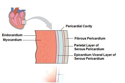
Starting from the inside to the outside the myocardium is the first layer of the heart and in turn is the thickest. The innermost layer of the cardiac wall is known as the endocardium. Its walls consist of of three tunics or layers.
The myocardium is the most significant of the three layers of the heart in terms of producing cardiac. Find step-by-step Anatomy and physiology solutions and your answer to the following textbook question. The wall of arteries and veins consists of following layers from inside to outside.
Describe the three layers of the heart. Three Layers of the Heart Wall. These tissue layers are highly specialized and perform different functions.
Describe the three major layers of the heart wall and how they relate to the pericardium. During ventricular contraction the wave of depolarization from the SA and AV nodes moves from within the endocardial wall through the myocardial layer to the epicardial. It is the most massive part of the heart.
Myocardium is the hearts muscular middle layer wall. Which of the three layers is most important in causing contractions of the heart. The endocardium is homologous with the intima of the blood vessels.
The heart wall itself can be divided into three distinct layers. It preforms the function of pumping what is necessary for the circulation of blood. The base muscle is considered because it is located in the inner part of the heart.
Epicardium -the outside of the myocardium is covered with a thin layer called the epicardium Visceral layer of serous pericardium. The heart wall is comprised of three layers the epicardium outer myocardium middle and endocardium inner. Identify and describe the function of the heart chambers major vessels that enter and exit each chamber and the atrioventricular valves semilunar valves chordae tendineae and papillary muscles.
Click card to see definition. Tap card to see definition. This is the middle layer of the heart that contains the cardiac muscular tissue.
Identify and describe the function of the heart chambers major vessels that enter and exit each chamber and the atrioventricular valves semilunar valves chordae tendineae and papillary muscles. The layers of heart wall is internal structure while pericardium is external structure of a heart. Tap again to see term.
The outermost layer is the epicardium which is derived from the proepicardium from the septum transversum. Identify the three layers of arteries and veins. Epicardium is the hearts outermost layer of protection.
It lines the cavities and valves of the heart. The endocardium myocardium and epicardium. Click again to see term.
The heart contains three basic layers similar to those seen in arteries and veins. This thin layer. The heart is composed of three layers.
Three layers of tissue form the heart wall. Describe the three major layers of the heart wall and how they relate to the pericardium. Epicardium myocardium and endocardium are the three layers that make up the heart wall.
Chambers of the Heart. The layers of the heart are as follows. Identify the three layers of arteries and veins.
Describe the three layers of the heart wall. The outer layer called pericardium Middle layer is made of myocardium Inner lining is called endocardium. These layers are called the endocardium myocardium and pericardium.
The epicardium outer layer which prevents excess expansion or movement of the heart the myocardium middle layer which initiates contractions driving the cardiac cycle and the endocardium inner layer that lines the cavities and valves. Myocardium middle layer Click card. Endocardium- This is the innermost layer and protects the valves and the heart.
The middle layer is the myocardium and the innermost layer is the endocardium which originated from mesothelial cells of the outflow tract. A muscular wall known as the septum separates the two sides of the heart. Myocardium- This layer forms the heart muscles.
Pericardium or the layer surrounding the heart Epicardium the outside layer of the heart Myocardium the middle layer of the heart Endocardium the innermost layer of the heart.
Anatomy Of The Human Heart Physiopedia

The Three Layers Of The Heart Wall Location Structure Function Video Lesson Transcript Study Com

Heart Anatomy Anatomy And Physiology I


0 Comments